It consists of a fluorophore which forms from the cyclization of the peptide bond and is used popularly as a biological marker. When GFP is employed as a tag in fusion with a protein, it has been found that it doesn't alter the protein's functioning or mechanisms in any way. which makes it suitable for both, the desired purpose i.e. to visualize the protein and the result i.e. successful fusion of GFP as a tag without producing any changes in the protein. GFP is extremely useful into looking at the inner workings of cells. All you have to do is introduce it into the desired environment and examine it under ultra violet light. The protein absorbs this ultra violet light and emits a low-energy green light. The application of GFP can be understood very simply by taking an example of a virus. When you tag a virus with GFP and infect a host with it, then under the effect of ultra violet light you can observe the virus propagating and invading the host's bosy just as by the green glow increases and spreads over the host's body.
In early 1960-70s, a scientist named Osamu Shimomura studied the GFP protein in addition to another protein called aequorin. These are the wild type GFP. When aequorin interacts with Ca2+ , fluorescence is produced by GFP which results in a blue color glow. A part of this energy produced by luminescence is transferred to GFP which leads to production of a green glow. However there are some GFP derivatives too which were produced as mutants as a result of genetic engineering. The picture below shows a jelly fish and the site of action for wild-type GFP & aequorin protein on it. The aequorin emits blue light which results in transfer of energy to GFP causing it to emit green light. The luminescence produced by aequorin results in
Ever since the study of biochemical and molecular mechanisms inside living beings started, a need was felt to examine these processes more closely so as to understand the life better. From the propagation of a disease-causing pathogen/molecule inside the body to development of a particular type of cell. How a cell initiates its functions inside body, how the developmental stages occur in a cell, how proteins fold and interact etc all come under this.
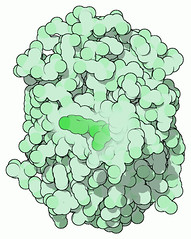
Of course an understanding of all these processes needed something so accurate that doesn't depend on other factors to regulate itself. GFP has revolutionized the branch of biological sciences by acting as a marker gene, reporter gene and a fusion tag. The green fluorescent protein or 'GFP' is a novel protein that is produced by the bio luminescent jellyfish Aequorea victoria. It doesn't require a co-factor and has also been used as an active indicator for protease action. The solutions of GFP appear yellow in room light but when exposed to sun light, they emit a bright green color. The image below has been taken from the RCSB data-bank for proteins and shows the structure of GFP.
It consists of a fluorophore which forms from the cyclization of the peptide bond and is used popularly as a biological marker. When GFP is employed as a tag in fusion with a protein, it has been found that it doesn't alter the protein's functioning or mechanisms in any way. which makes it suitable for both, the desired purpose i.e. to visualize the protein and the result i.e. successful fusion of GFP as a tag without producing any changes in the protein. GFP is extremely useful into looking at the inner workings of cells. All you have to do is introduce it into the desired environment and examine it under ultra violet light. The protein absorbs this ultra violet light and emits a low-energy green light. The application of GFP can be understood very simply by taking an example of a virus. When you tag a virus with GFP and infect a host with it, then under the effect of ultra violet light you can observe the virus propagating and invading the host's body just as by the green glow increases and spreads over the host's body.
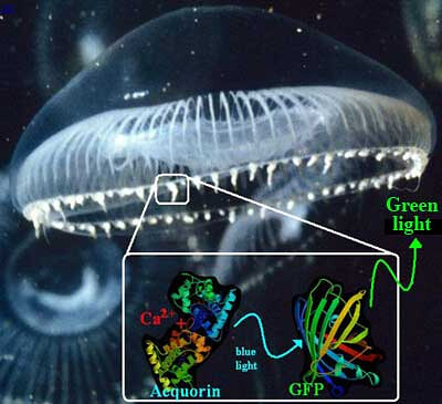
In early 1960-70s, a scientist named Osamu Shimomura studied the GFP protein in addition to another protein called aequorin. These are the wild type GFP. When aequorin interacts with Ca2+ , fluorescence is produced by GFP which results in a blue color glow. A part of this energy produced by luminescence is transferred to GFP which leads to production of a green glow. However there are some GFP derivatives too which were produced as mutants as a result of genetic engineering. The picture below shows a jelly fish and the site of action for wild-type GFP & aequorin protein on it. The aequorin emits blue light which results in transfer of energy to GFP causing it to emit green light. The luminescence produced by aequorin results in emission of energy, some of which is transferred to GFP here.

The first GFP derivative was reported back in 1995 by Rogen Tsein, which had a point mutation S65T. This mutant GFP had better characteristics than the wild type GFP; the spectral properties were found to have improved in the mutated GFP leading to an increased fluorescence. Ever since then many genetic engineering experiments have been performed on GFP and many other type of mutant derivatives have been produced, for example: color mutants which mainly include blue fluorescent protein (namely Azurite, EBFP2 etc.), yellow fluorescent protein (namely Citrine, Venus etc.); roGFPs which are the redox sensitive version of GFP and are engineered by introduction of cysteine molecules etc.
Other than serving as a cell marker, GFP is also used widely for protein localization by the researchers. When the fluorescent feature of GFP is combined with con-focal microscopy, it helps in determining nuclear localization of polypeptides. Researchers have also produced green mice by incorporating the GFP producing gene in mice.
The use of GFP is expanding in the field of arts too. Genetically engineered fishes, rabbits, plants, rats, mice, frogs, flies, worms have been produced that glow green in color (or any other color mutant form of GFP, for example blue fluorescent protein) with the application of GFP. Sure, the use of these genetically modified organisms may raise issues as long as ethics are concerned.
It wouldn't be an exaggeration to say that the discovery of GFP protein has revolutionized the field of bio-medical science. Molecules can be tagged with GFP in order to track their growth and activity either in-vivo or in-vitro. GFP has been successfully used as a marker and a tag in several of experiments, for example- in an experiment, the sperm of fruit fly was tagged with GFP to see it in action and how the fertilization was carried out, scientists attached GFP with insulin producing cells, in pancreas, to see their functioning. In many of the experiments, utilizing the same methods can give give insights onto how a disease is developed by tracking the development of the disease causing molecule or cells and their growth and effective measures can then be successfully developed to inhibit the growth.
Thus it can be concluded and put in one line that GFP has provided the fields of biological sciences an important tool to understand the mechanisms of life and the not so beneficial mechanisms which interrupt the normal functions of cells which has provided a better insight into dealing with diseases and developing therapeutic strategies accordingly. of energy, some of which is transferred to GFP here.
The first GFP derivative was reported back in 1995 by Rogen Tsein, which had a point mutation S65T. This mutant GFP had better characteristics than the wild type GFP; the spectral properties were found to have improved in the mutated GFP leading to an increased fluorescence. Ever since then many genetic engineering experiments have been performed on GFP and many other type of mutant derivatives have been produced, for example: color mutants which mainly include blue fluorescent protein (namely Azurite, EBFP2 etc.), yellow fluorescent protein (namely Citrine, Venus etc.); roGFPs which are the redox sensitive version of GFP and are engineered by introduction of cysteine molecules etc.
Other than serving as a cell marker, GFP is also used widely for protein localization by the researchers. When the fluorescent feature of GFP is combined with con-focal microscopy, it helps in determining nuclear localization of polypeptides. Researchers have also produced green mice by incorporating the GFP producing gene in mice.
The use of GFP is expanding in the field of arts too. Genetically engineered fishes, rabbits, plants, rats, mice, frogs, flies, worms have been produced that glow green in color (or any other color mutant form of GFP, for example blue fluorescent protein) with the application of GFP. Sure, the use of these genetically modified organisms may raise issues as long as ethics are concerned.
It wouldn't be an exaggeration to say that the discovery of GFP protein has revolutionized the field of bio-medical science. Molecules can be tagged with GFP in order to track their growth and activity either in-vivo or in-vitro. GFP has been successfully used as a marker and a tag in several of experiments, for example- in an experiment, the sperm of fruit fly was tagged with GFP to see it in action and how the fertilization was carried out, scientists attached GFP with insulin producing cells, in pancreas, to see their functioning. In many of the experiments, utilizing the same methods can give give insights onto how a disease is developed by tracking the development of the disease causing molecule or cells and their growth and effective measures can then be successfully developed to inhibit the growth.
Thus it can be concluded and put in one line that GFP has provided the fields of biological sciences an important tool to understand the mechanisms of life and the not so beneficial mechanisms which interrupt the normal functions of cells which has provided a better insight into dealing with diseases and developing therapeutic strategies accordingly.
About Author / Additional Info: